'Doing Science': the Broadened Scope of Undergraduate Cell Biology at UK
LEXINGTON, Ky. (April 23, 2015) — The realm of science in the United States — education, research and career opportunities — is always a hot topic, but especially so in the last several years. Technology has transformed students' learning experiences and the National Science Board (NSB) called on education and policy to foster "the next generation of STEM innovators."
In 2010, the University of Kentucky Department of Biology responded with a curriculum reform, changing the way undergraduate biology is taught at UK, and perhaps leading to more UK students pursuing scientific careers.
The curriculum reform, led by Vincent Cassone, department chair and professor, implemented new laboratory experiences in genetics, evolution, cell biology, physiology and ecology. To do that, the department had to renovate classroom space in the Thomas Hunt Morgan Biological Sciences Building and the Multidisciplinary Science Building into laboratories. The undergraduate cell biology lab was one of these.
Part of the Intro to Cell Biology course, or BIO 315, designed by Professor Rebecca Kellum and Lab Coordinator Seth Jones, the lab promotes hands-on, real-world research and experimentation.
As Cassone says, "science is something you do, and biology is a physically active scientific exercise," and a high-tech lab was needed to put classroom concepts to practice, especially for cell biology, known to be an abstract discipline for students.
But the lab at UK took the undergraduate experience one step further than most by updating standard microscopes to fluorescence microscopy. Cells can only be seen through microscopes, and molecules carrying out their functions must be labeled with fluorescence in order to view them through the microscope, but it is unusual to perform that level of microscopy for undergraduates.
"Labs at almost all universities are equipped with standard light microscopes, but few are equipped with fluorescent microscopes. If they are, it is usually limited to a single shared instrument for the whole lab," said Kellum, who began teaching cell biology at UK in 1999.
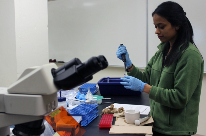
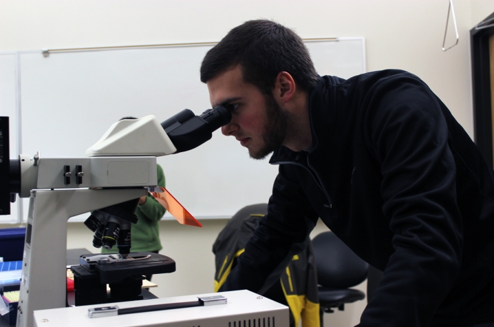
The UK undergraduate cell biology lab instead features a fluorescent microscope for each pair of lab partners in course sections of 30 students.
"We have found that students are only interested in looking at specimens prepared by their own hands on their own microscopes," Kellum said.
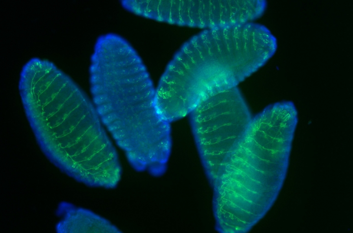
Students produce their own images of nanoparticles normally out of sight to the human eye, such as those below featuring drosophila embryos with immunofluorescence of a neuronal protein (green) and DAPI (a fluorescent stain) (blue). That particular lab activity is typically not well suited for undergraduate labs because it requires overnight incubations and fine precision, Jones said. To ensure UK students could experience the activity, Kellum developed protocol to get the immunofluorescence technique working within a single three-hour lab period.
But learning microscopy methods and producing cellular images that could double as artwork isn't the entire scope of the lab course. It also includes engaging in actual research, and learning how science works in general — forming hypotheses, testing them, finding out if results support the hypotheses or not.
Following the lab, teaching assistants guide students in using their creativity to plan a set of hypothetical experiments using those techniques and other fluorescent labeling techniques.
"They get the flavor of real-world research, they get to do cellular and molecular biology related techniques here that they study in their lecture materials. And they need to do experiments to find out if things work the way they think they do, or not," said Swagata Ghosh, a cell biology lab teaching assistant and doctoral student in biology.
"When they see proteins glowing, when they see proteins on a gel or they look at DNA under UV light — maybe things they've just read about in their textbooks — that gives them more encouragement or interest in studying the subject," Ghosh said.
Another experiment performed by undergraduates in the lab is western blotting, a technique that separates and identifies proteins within a sample of tissue, and in their case, chicken breast muscle tissue. Students also use centrifugation to isolate mitochondria from chicken liver, from which they measure the rate of reaction of an enzyme involved in cellular respiration.
Jones says students also dissect and view the giant polytene chromosomes from the salivary glands of fruit fly larvae, re-enacting a technique that allowed UK alumnus Thomas Hunt Morgan to identify the chromosome as the location of genes.
"The experience that those students are getting in cell biology, now taught by Drs. Ed Rucker, Rebecca Kellum and Seth Jones, really is a top-notch experience," Cassone said.
And the results prove it — class performance on exams has improved by at least a letter grade, and the drop, fail and withdrawal rate has become virtually non-existent.
With the development of the neuroscience degree and plans for a new multidisciplinary research facility at UK, experiences in biology — and science education and research overall — at the university will continue to evolve, producing tomorrow's "STEM innovators" right on campus.
MEDIA CONTACT: Whitney Harder, 859-323-2396, whitney.harder@uky.edu


