


UK HealthCare pioneers emergency department ultrasound program for vision-threatening disease
LEXINGTON, Ky. (June 29, 2026) — A new initiative at the University of Kentucky is utilizing cutting-edge bedside imaging technology in the UK Albert B. Chandler Hospital Emergency Department with the goal of diagnosing a serious, vision-threatening condition faster — and potentially preventing irreversible blindness.
The new program stems from years of work by Jagannadha “Jay” Avasarala, M.D., Ph.D., neurologist with Kentucky Neuroscience Institute and professor of neurology in the UK College of Medicine. His latest study on the subject, published in the American Journal of Emergency Medicine, revealed a critical blind spot in how emergency departments across the globe diagnose a condition known as giant cell arteritis (GCA), which is blood vessel inflammation that can suddenly block blood flow to the eye. The findings revealed that many patients at or over 50 who arrive in emergency departments with sudden vision changes are not tested for this potentially blinding disease.
Because of these findings the team at UK is launching a pioneering point-of-care Temporal Artery Ultrasound (TAUS) Program for evaluating GCA. The initiative marks what doctors believe is the first program in the country to integrate real-time bedside temporal artery ultrasound into emergency department care led in part by neurology residents.
Although GCA, sometimes referred to as a “stroke of the eye,” is considered a medical emergency by both the American Stroke Association and the American Academy of Ophthalmology, Avasarala said there are currently no emergency department guidelines requiring hospitals to track or rapidly evaluate these cases using this type of imaging.
“The diagnosis of giant cell arteritis affecting the eye as an acute problem is an emergency,” Avasarala said. “We are taking this up because rapid diagnosis and treatment are critical to preventing blindness.”
Traditionally, diagnosing GCA has often relied on temporal artery biopsies or delayed outpatient imaging appointments. The new TAUS program at UK instead brings ultrasound directly to the bedside in the emergency department, allowing providers to quickly identify the “halo sign,” a hallmark indicator of vessel wall inflammation associated with the disease.
That speed can make a significant difference for patients.
“Rapid and immediate diagnosis and treatment are essential,” Avasarala said. “This is done to prevent blindness.”
Studies have shown that vision loss can occur in up to 20% of patients with giant cell arteritis worldwide if the condition is not identified and treated quickly.
The initiative builds on the growing use of point-of-care ultrasound, commonly known as POCUS, across emergency medicine and other specialties. At UK, emergency medicine and neurology physicians are collaborating to apply the technology to ischemic ophthalmic emergencies in a new way.
Evan Vincent, M.D., program director for UK College of Medicine’s Emergency Ultrasound Fellowship, said point-of-care ultrasound has transformed the ability of physicians to diagnose conditions quickly at the bedside.
“Using ultrasound to examine the temporal artery is a newer approach that has been studied but is not yet routine for eye-related emergencies,” Vincent said. “We are excited to use this bedside technology to identify this serious, vision-threatening condition more quickly and to work closely with our neurology colleagues and trainees to improve patient care.”
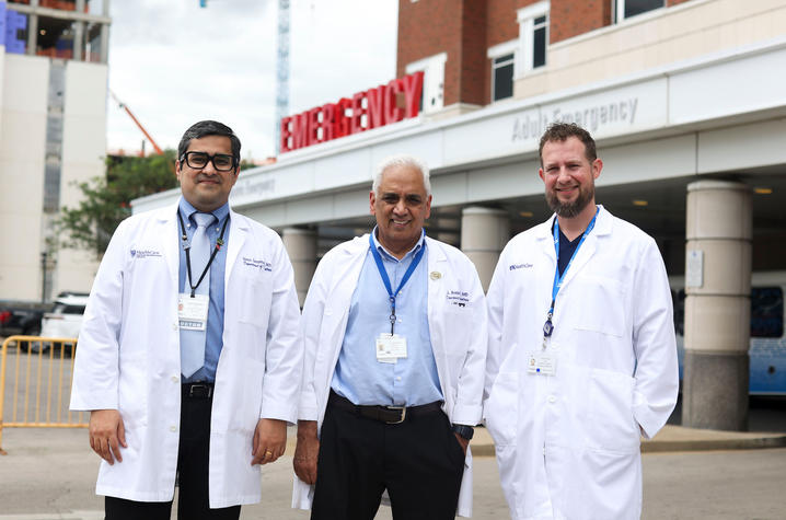
The new program is led by Suhas Gangadhara, M.D., UK College of Medicine assistant professor of neurology, with support from Vincent and UK’s emergency medicine ultrasound experts. In addition to improving patient care, the initiative also creates a unique educational opportunity for neurology residents, who will help perform and interpret the ultrasounds in emergency settings.
“There is no program like this across the country, certainly not from a resident’s perspective, let alone from the emergency department perspective,” said Gangadhara. “Our residents are learning a new technique and will be at the forefront of this project.”
Providers hope the initiative will ultimately help establish a new national standard for diagnosing vascular emergencies affecting the eye — one centered on faster bedside evaluation, quicker treatment and better outcomes for patients at risk of losing their vision.
UK HealthCare is the hospitals and clinics of the University of Kentucky. But it is so much more. It is more than 10,000 dedicated healthcare professionals committed to providing advanced subspecialty care for the most critically injured and ill patients from the Commonwealth and beyond. It also is the home of the state’s only National Cancer Institute (NCI)-designated Comprehensive Cancer Center, a Level IV Neonatal Intensive Care Unit that cares for the tiniest and sickest newborns and the region’s only Level 1 trauma center.
As an academic research institution, we are continuously pursuing the next generation of cures, treatments, protocols and policies. Our discoveries have the potential to change what’s medically possible within our lifetimes. Our educators and thought leaders are transforming the healthcare landscape as our six health professions colleges teach the next generation of doctors, nurses, pharmacists and other healthcare professionals, spreading the highest standards of care. UK HealthCare is the power of advanced medicine committed to creating a healthier Kentucky, now and for generations to come.