Light Microscopy Core Instruments, Staff Boost Research Capacity at UK

LEXINGTON, Ky. (Nov. 16, 2016) — The Light Microscopy Core, a newly named research core facility under the auspices of the Office of the Vice President for Research, has invested $1.3 million in two new microscopes to support an array of research across the University of Kentucky.
Chris Richards, director of the Light Microscopy Core and assistant professor of chemistry, said these instruments and the hiring of manager Thomas Wilkop will enable UK researchers to utilize the most advanced imaging available.
“If we want to understand biological systems, ranging from neuroscience to physiology, or apply imaging techniques for cutting-edge materials science, we really need to have the type of equipment that makes us competitive with other universities. These microscopes absolutely do it,” said Richards.
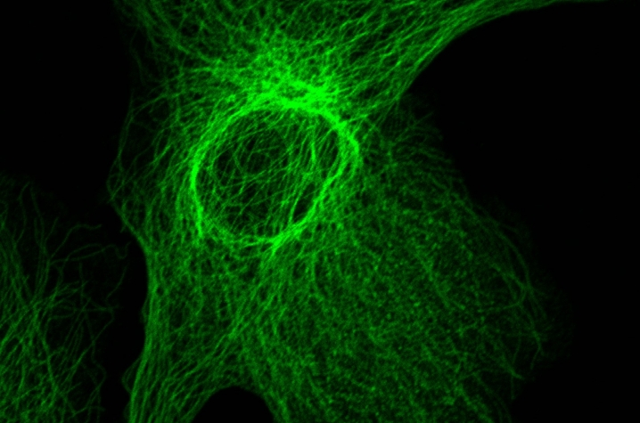
The first new microscope, a Nikon A1RSi, is already available to researchers. “This instrument is a confocal microscope with enhanced resolution,” Richards said. “Normally, with a confocal microscope you can achieve a resolution of 200 to 225 nanometers. This one allows you to reach 150 to 180 nanometers. It can give you much better spatial resolution, so you can see finer features. You can also get much better temporal resolution — meaning you can go much faster than with a typical confocal microscope.”
The second new microscope is the Nikon SIM/STORM. UK is hosting a training seminar on this instrument, led by Nikon, at 1:30 p.m. Monday, Nov. 21, in 202A BBSRB (Biomedical Biological Sciences Research Building). This seminar is open to anyone who is interested.
“One feature of this super resolution microscope, called STORM, can theoretically get us down to about 30 nanometer resolution. Beyond the beautiful images, you can quantify which proteins are interacting with each other or determine if interacting proteins are in the same organelle or subcellular compartment within a cell,” Richards said. “A second feature on that microscope is called structured illumination microscopy, SIM for short, which has a resolution of about 85 nanometers. The nice thing about the SIM part is you can do it on a live cell. SIM can actually allow you to image live cells for hours if you want. This gives you the ability to track subcellular motion and dynamics over time.”
Richards said the Light Microscopy Core will offer free training, lasting roughly an hour to an hour and a half depending on past imaging experience, to UK researchers interested in using these microscopes. That training will be led by Thomas Wilkop, the new manager of the Light Microscopy Core since Nov. 15, 2016.
Wilkop, who came to UK from the University of California, Davis, has a doctoral degree in electronics engineering from Sheffield Hallam University in Sheffield, United Kingdom. He previously served as the manager of a single molecule detection facility, and his most recent project involved using fluorescence microscopy to study plant metabolism. He has extensive experience in developing and building new microscopy instruments, developing new methodologies and applying quantitative image analysis tools.
For more on the Light Microscopy Core, visit www.research.uky.edu/core/imaging.


