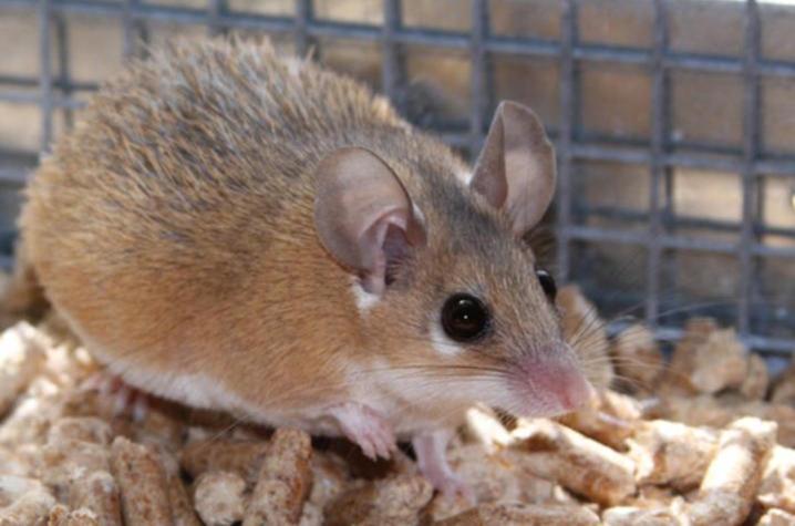
Do spiny mice hold the key to regenerative healing? UK study explores

LEXINGTON, Ky. (April 26, 2023) — Research at the University of Kentucky is looking to nature to better understand cellular processes that permit lost tissue to regenerate in spiny mice — processes that might lie dormant in humans.
Ashley Seifert, Ph.D., an associate professor in the UK College of Arts and Sciences’ Department of Biology, teamed up with scientists in Germany and the Netherlands to tease apart how identical injuries in two different rodent species lead to regenerative healing in one case but not the other.
The paper, published today in Science Advances, compared tissue healing in laboratory mice and spiny mice. Whereas adult laboratory mice heal injuries with scar tissue, Seifert’s research group has pioneered using spiny mice to understand how complex tissue can regenerate in a mammal. Spiny mice are known for their unique ability to regrow lost skin, restore function to a severed spinal cord and, as one of Seifert’s previous studies shows, repair damaged heart tissue to effectively maintain heart function.
“Over the last decade, spiny mice have emerged as a key animal model that can bridge regenerative biology and medicine,” said Seifert.
“Injury response of most human organs yields fibrotic tissue resulting in permanent scars and organ function failure,” said Antonio Tomasso, lead author on the study and a Ph.D. student at the Max Planck Institute for Molecular Biomedicine in Germany. “Our study investigates the diverging mechanisms driving fibrosis and regeneration in adult mammals and sets the stage to develop new therapeutic strategies aiming to repair and regenerate damaged organs.”
Tomasso had studied extracellular signal-regulated kinase (ERK) activity in planarian flatworms with Kerstin Bartscherer, Ph.D., research group leader at the Max Planck Institute, and subsequently built a strong collaboration with UK’s Seifert after his pioneering studies on tissue regeneration in spiny mice.
A series of experiments were performed on spiny and lab mice at UK and the Hubrecht Institute in the Netherlands. Researchers tracked the cellular response to injury during tissue healing and noted that ERK-mediated signaling acts as a key switch controlling the healing process.
Researchers observed ERK activity in response to tissue damage in both rodent species. However, as the initial wound healing phase and tissue inflammation resolved, ERK activity dropped off in lab mice. In contrast, ERK-signaling was maintained in a diverse array of cell types at high levels in spiny mice as new tissue was generated.
As part of the study, the researchers applied ERK activators to the ear of lab mice and were able to generate the same type of regenerative healing they observed in spiny mice. Researchers found that sustained, high ERK activity is critical for regeneration. That partially explains why some closely related species, like spiny mice and lab mice, may have very different cellular responses to the same injury.
“Findings from our study suggest that sustaining ERK-signaling beyond the initial wound healing phase promotes proliferation over collagen production. It also appears that ERK acts as a type of cellular sensor, similar to how a sound engineer controls levels at a mixing board,” said Seifert. “While ERK is unlikely to be the only sensor, our results show that increasing its activity in an injury can antagonize fibrosis and promote regeneration.”
“In our research, we also asked whether poor regenerators, namely laboratory mice and humans, have completely lost their ability to regenerate or whether they still possess latent regenerative traits. If the latter hypothesis holds true, it would have enormous implications for regenerative medicine, implying that a regenerative response can still be triggered in cells and tissues of poorly regenerating adult mammals and, ultimately, humans,” said Tomasso.
Researchers say more studies are needed to determine if the regeneration of other organs or digits can be accomplished.
This team of researchers represents UK, the Max Planck Institute for Molecular Biomedicine and Osnabrück University in Germany, the Royal Netherlands Academy of Arts and Sciences, and the Princess Máxima Center for Pediatric Oncology in the Netherlands.
Research reported in this publication was supported by the National Institute of Arthritis and Musculoskeletal and Skin Diseases of the National Institutes of Health under Award Number R01AR070313 and the National Institute of Dental and Craniofacial Research of the National Institutes of Health under Award Number R21DE028070. The content is solely the responsibility of the authors and does not necessarily represent the official views of the National Institutes of Health.
Research reported in this publication was supported by the National Science Foundation under Award Number 1353713. The opinions, findings, and conclusions or recommendations expressed are those of the author(s) and do not necessarily reflect the views of the National Science Foundation.
As the state’s flagship, land-grant institution, the University of Kentucky exists to advance the Commonwealth. We do that by preparing the next generation of leaders — placing students at the heart of everything we do — and transforming the lives of Kentuckians through education, research and creative work, service and health care. We pride ourselves on being a catalyst for breakthroughs and a force for healing, a place where ingenuity unfolds. It's all made possible by our people — visionaries, disruptors and pioneers — who make up 200 academic programs, a $476.5 million research and development enterprise and a world-class medical center, all on one campus.


